ULTRASOUND IN FACIAL AESTHETICS
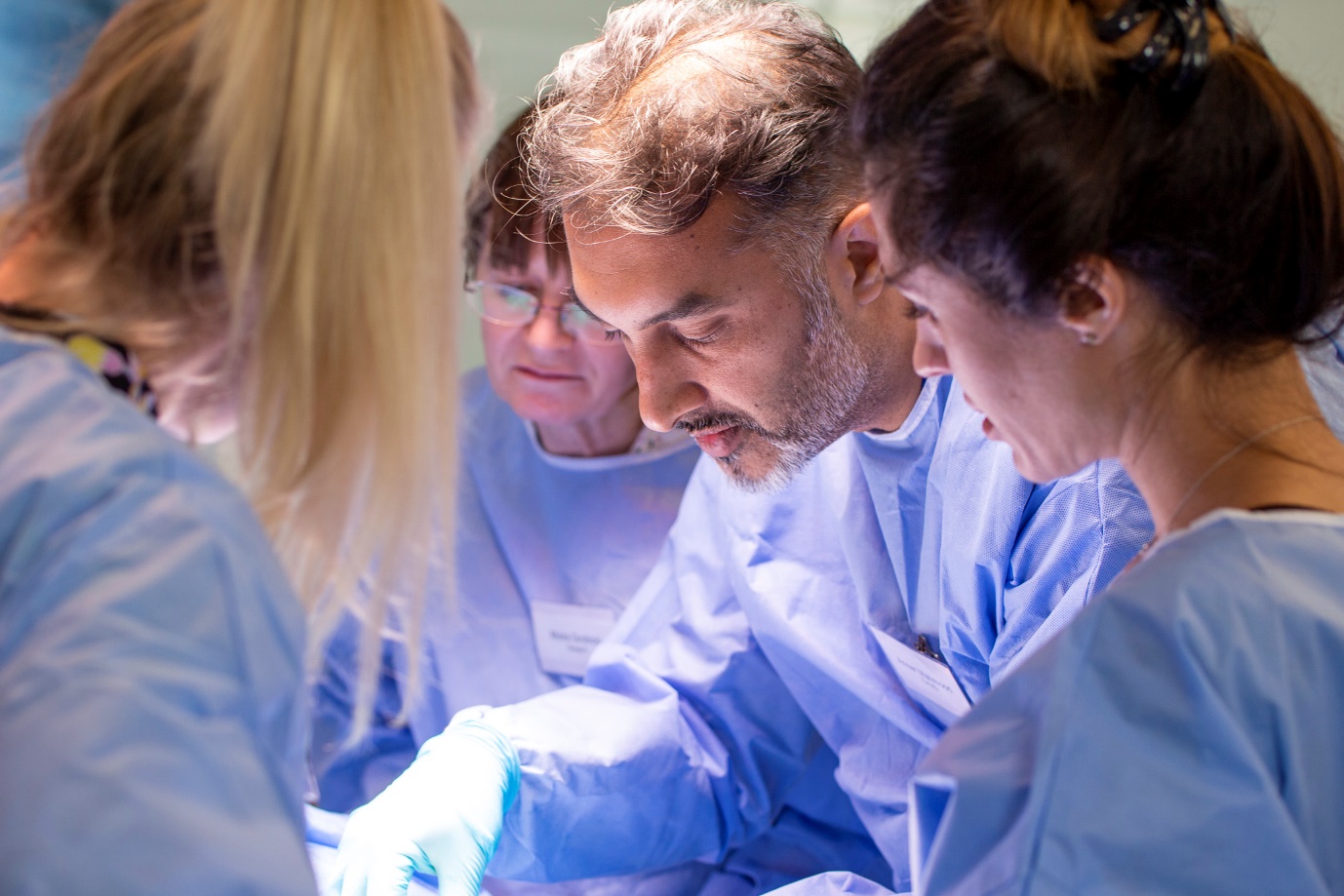
Consultant Surgeon Mr Ansar Mahmood explains why he believes aesthetic clinicians will be routinely using ultrasound in medical aesthetic practice
A sound footing in anatomy is crucial to minimise risk and optimise outcomes in many aspects of interventional medicine; the same applies to facial aesthetic injections. However, it is also well recognised that there is significant variation between individuals. This is most important in practical terms when concerning the arterial tree.
Ultrasound can give you important insight into variations in facial anatomy, particularly with the correct training and with regular practice. An ultrasound device consists of a probe and a processor. The probe will generate a sound wave that penetrates body tissue. Sound waves interact with the tissue and become progressively weaker in strength as the waves are absorbed or scattered.
Part of the sound waves is being reflected. The reflected sound waves, picked up by the probe and directed to the processor, are transformed into a digital image.
When using ultrasound, you not only increase the precision of your treatments by ensuring the correct anatomical layer is targeted, but you also increase the safety of injectable treatments by visualising important structures and avoiding them.
The stated normal variation in anatomy from client to client makes it impossible to be safe every time; the simple rule applies in all walks of life – ‘that every action or intervention generates risk’ – this includes crossing the road, kicking a ball, or using an injection. It is impossible to completely avoid complications or sub-optimal placement of injectables, even in the most experienced clinicians’ hands.
This risk is not completely removed by ultrasound but narrows the variance in practice and improves assessment and targeting to allow precision placement of product. I have been using ultrasound in high-risk clinical areas or where precision delivery of the product is key for over five years and have seen where a 1mm change in placement in my needle has avoided perforation of an artery.
It is well recognised, however, that becoming adept like most skills requires ongoing practice, and there is a steep learning curve initially. My children and other family members were often subjected to being scanned, often while trying to watch television, when I started my ultrasound journey!
There are some excellent ‘phantoms’ that can be used to practice safely and away from patients, particularly for targeting and avoiding anisotropy – where the beam of the ultrasound is tangential to the structure being looked at, rather than perpendicular, and give artefact. It is very important to recognise normal so that you can recognise abnormal readily when you see it.
In complications management, in particular for hyaluronic acid- based fillers, it has become routine practice for me and those trained in ultrasound use to depend on precision recognition of placement and targeted removal with or without the use of hyaluronidase (Hyalase®). This not only allows a more targeted removal but also facilitates the use of much smaller doses of Hyalase, hence reduces the risk of damage to the patient’s native collagen.
I am certain ultrasound targeting will become the standard of care in aesthetics in high-risk areas and almost certainly in complications management in the not-too-distant future. The availability in recent years of high-resolution, portable ultrasound scanners that, from a cost perspective, are accessible to most clinics will hopefully lead to the rapid adoption of this game-changing technology.
The digital platforms that have been created allow remote supervision and continuing professional development with mentorship readily adoptable.
I am passionate about the scientific endeavour and optimising patient safety in our practice of medicine and surgery and hope to bring this philosophy to the forefront in our training courses. My vision is that this will assist in producing evidence-based protocols for our pioneering treatments while providing reassurance to patients that they are being treated with the latest and safest advances.
Mr Ansar Mahmood
Mr Ansar Mahmood is a Consultant Surgeon in one of the leading Major Trauma units in the UK. Fellowship Trained in Orthopaedic Trauma Surgery. Research interests include causes of cell death and ischaemia, Platelet Rich Plasma (PRP), cell therapy and Orthobiologics and their use in musculoskeletal conditions, tissue regeneration and wound healing. He founded the Academy of Regenerative Medicine in the UK.
For PDF Version click here: ULTRASOUND IN FACIAL AESTHETICS – Consultant Surgeon Mr Ansar Mahmood (1)
- A detailed observation of variations of the facial artery, with emphasis on the superior labial artery. Surg Radiol Anat. 2006 Jun;28(3):316-24. doi: 10.1007/s00276-006-0093-0
- The utility of high-frequency ultrasoundin dermal filler evaluation. Ann Plast Surg.2011 Nov;67(5):469-73. doi: 10.1097/SAP.0b013e318203ebf6